
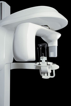
3D Imaging
Cone Beam CT Scan
Our philosophy is always directed toward evidence-based patient care with the safety of our patients of foremost importance. Although the newest technologies have made radiation exposure extremely low, we strive to expose patients to the least amount of radiation necessary for the greatest amount of diagnostic information. For cases where 3-dimensional localization of teeth or anatomical structures are of significance, cone beam CT imaging is an extraordinarily important advancement to provide the highest level of care possible.
Cary Oral Surgery was one of the pioneers in Cone Beam CT imaging for oral and maxillofacial surgical care. We were one of the first in North Carolina to incorporate this technology to provide the safest and highest level of care possible. We recently introduced our second cone beam unit to provide the highest level of detailed imagery while allowing for even lower radiation exposures.
The CS 9300 system gives our oral and maxillofacial practice state-of-the-art technology to help Dr. Vande Berg and Dr. Englehardt diagnose potential issues more accurately and provide treatment with unprecedented confidence. Unlike a traditional spiral CT scanner, this 3D system utilizes cone beam CT technology and provides precise, crystal-clear digital images while minimizing your exposure to radiation.
The CS 9300 scanner allows us to use less radiation and often can choose the field of view, or scanning area, that best suits your specific treatment needs. This helps to limit your radiation exposure because we are focusing specifically on your area of concern.
The CS 9300 system brings the latest 3D technology to Cary Oral & Maxillofacial Surgery, providing unmatched visualization of anatomical detail to aid in treatment planning and helps us to better explain the particulars of your case as well as address any questions you may have. Drs. Vande Berg and Englehardt can use this innovative technology to quickly and easily share 3D images of the area of concern with your referring doctor – allowing the doctors to collaborate on your care, improving your experience, and delivering a positive treatment outcome.